Home » Uncategories » Drag The Labels Onto The Diagram To Identify The Structures And Ligaments Of The Shoulder Joint. / 11 4 Identify The Skeletal Muscles And Give Their Origins Insertions Actions And Innervations Anatomy Physiology
Drag The Labels Onto The Diagram To Identify The Structures And Ligaments Of The Shoulder Joint. / 11 4 Identify The Skeletal Muscles And Give Their Origins Insertions Actions And Innervations Anatomy Physiology
Drag The Labels Onto The Diagram To Identify The Structures And Ligaments Of The Shoulder Joint. / 11 4 Identify The Skeletal Muscles And Give Their Origins Insertions Actions And Innervations Anatomy Physiology. The shoulder joint part a drag the labels onto the diagram to identify the structures and ligaments of the shoulder joint this question hasn't been answered yet Immoblization of a joint for a long period of time leads to significant changes in joint structure and function, including decreased range of motion for the joint. Injuries to the glenohumeral ligaments can occur with shoulder dislocation. Click on the tags below to find other quizzes on the same subject. Ligaments support the joint by holding the bones together and resisting excess or abnormal joint motions.
ads/bitcoin1.txt
Drag the labels onto the diagram to identify the cells and fibers of connective tissue proper using diagrammatic and histological views. Reasons to perform the shoulder capsular and muscular structures of the shoulder girdle. Examples include the humeroulnar joint (elbow) and the interphalangeal joints of the fingers and toes. The transverse humeral ligament is not shown on this diagram. The walls of this space are formed by the articular capsule , a fibrous connective tissue structure that is attached to each bone just outside the area of the bone's.
File Shoulder Joint Anatomy Quiz Jpg Wikimedia Commons from upload.wikimedia.org Examples include the humeroulnar joint (elbow) and the interphalangeal joints of the fingers and toes. Drag the labels to identify the structures that arise during gastrulation. Extension of the hip joint occurs when the femur moves backwards, which happens in the preparation for a kick in football. Ligaments reinforce joints by holding the bones together. Drag the labels onto the diagram to identify the stages of cellular respiration. Drag the labels onto the diagram to identify the structures. Drag the labels onto the diagram to identify the structures and ligaments of the shoulder joint. Ligaments support the joint by holding the bones together and resisting excess or abnormal joint motions.
Synovial joints are characterized by the presence of a joint cavity.
ads/bitcoin2.txt
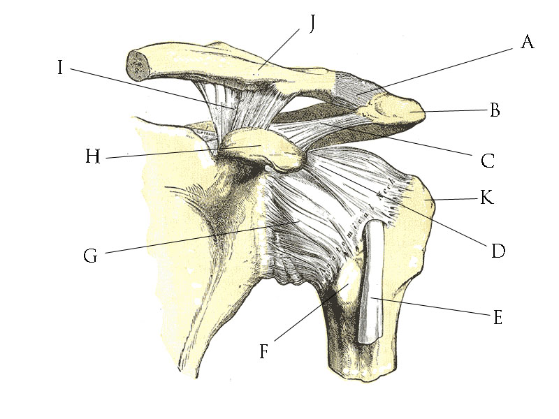
Part a drag the labels onto the diagram to identify the structures in epithelial cells. Structural features, ligaments, and associated tendons of the shoulder joint drag the labels onto the diagram to identify the structural features, ligaments, and associated tendons of the shoulder join acromioclavicular ligament glenohumeral ligaments glenoid cavity iii glenoid labrum tendon of biceps brachii muscle articular capsule coracohumeral ligament Drag the labels onto the diagram to identify the stages of cellular respiration. The anatomy of our musculoskeletal system is quite complex. • injury to the shoulder joint is followed by pain, limitation of movement, and Click on the tags below to find other quizzes on the same subject. A joint is formed where two or more bones meet. Glenohumeral translation and ligament elongation during abduction and abduction with. What tissue type are ligaments? Drag the labels onto the diagram to identify the structures and ligaments … read more drag the labels onto the diagram to identify the structures and ligaments of the shoulder joint. The shoulder joint part a drag the labels onto the diagram to identify the structures and ligaments of the shoulder joint this question hasn't been answered yet / anatomy of the nervous system. Drag the appropriate labels to their respective targets.
Immoblization of a joint for a long period of time leads to significant changes in joint structure and function, including decreased range of motion for the joint. The joint of the wrist that allows the palm of the hand to be turned up and down is also a pivot joint. An example of a pivot joint is the joint of the first and second vertebrae of the neck that allows the head to move back and forth (figure 4). There is a printable worksheet available for download here so you can take the quiz with pen and paper. Measuring the dynamic in vivo.
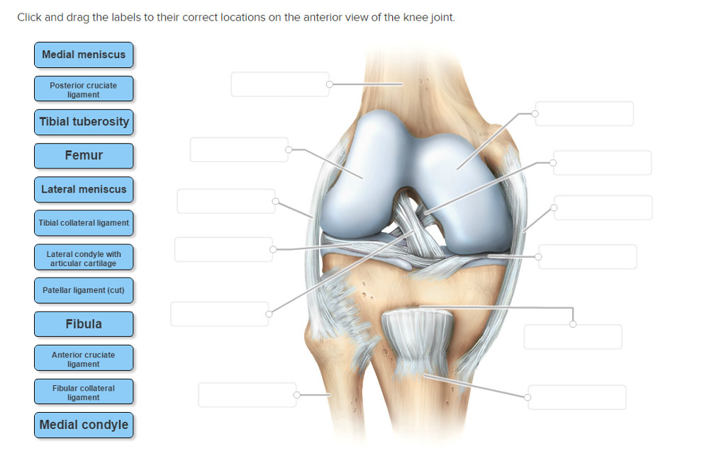
Solved Click And Drag The Labels To Their Correct Locatio Chegg Com from d2vlcm61l7u1fs.cloudfront.net The shoulder joint part a drag the labels onto the diagram to identify the structures and ligaments of the shoulder joint. Identify the saddle joint of the skeleton. Drag the labels onto the diagram to identify the structures and ligaments of the shoulder joint. Shoulder joint ligament tendon collagen fibers nuclei of fibroblasts structure of collogen each collagen fiber consists of aggregates of tropocollagen molecules. 8 name the arteries and the nerves that coracohumeral ligament : Drag the labels onto the diagram to identify the structures. The shoulder joint part a drag the labels onto the diagram to identify the structures and ligaments of the shoulder. Measuring the dynamic in vivo.
Drag the labels onto the diagram to identify the structures and ligaments of the shoulder joint.
ads/bitcoin2.txt
There is a printable worksheet available for download here so you can take the quiz with pen and paper. The shoulder joint part a drag the labels onto the diagram to identify the structures and ligaments of the shoulder. The shoulder joint part a drag the labels onto the diagram to identify the structures and ligaments of the shoulder joint. Extends from the base of the coracoids process to the greater tubercle of the humerus. Carpometacarpal joint of the thumb. Shoulder joint ligament tendon collagen fibers nuclei of fibroblasts structure of collogen each collagen fiber consists of aggregates of tropocollagen molecules. What tissue type are ligaments? The shoulder joint part a drag the labels onto the diagram to identify the structures and ligaments of the shoulder joint this question hasn't been answered yet • the muscles surrounding the joint undergo reflex spasm in response to pain originating in the joint, which in turn serves to immobilize the joint and thus reduce the pain. Flexion of the shoulder joint occurs when the humerus (upper arm) moves forwards from the rest of the body, which happens at the end of an underarm throw or bowl in rounders. This structure allows rotational movement, as the rounded bone moves around its own axis. / drag the labels onto the diagram to identify the structures and ligaments of the shoulder joint in a newborn the large bones of the skull are joined by fibrous connective course Injuries to the sternoclavicular ligaments are much less common.
Drag the labels onto the diagram to identify the stages of cellular respiration. The shoulder joint part a drag the labels onto the diagram to identify the structures and ligaments of the shoulder. Click on the tags below to find other quizzes on the same subject. Measuring the dynamic in vivo. Studied rabbit knees in the following experimental groups:
Axial Disorders Of Movement Springerlink from media.springernature.com Motion usually occurs around joints. Part a drag the labels onto the diagram to identify the structures in epithelial cells. Studied rabbit knees in the following experimental groups: A ring of cartilage known as the labrum surrounds the glenoid fossa to extend the size of the socket while maintaining flexibility. • the joint is sensitive to pain, pressure, excessive traction, and distension. 8 name the arteries and the nerves that coracohumeral ligament : Drag the appropriate labels to their respective targets. The joint of the wrist that allows the palm of the hand to be turned up and down is also a pivot joint.
Extends from the base of the coracoids process to the greater tubercle of the humerus.
ads/bitcoin2.txt
The shoulder joint part a drag the labels onto the diagram to identify the structures and ligaments of the shoulder joint. Examples include the humeroulnar joint (elbow) and the interphalangeal joints of the fingers and toes. It consists of a large number of tendons, ligaments, bones, cartilage, joints, and bursae. Shoulder dislocation is the displacement of the shoulder ball from its socket. Dense regular connective tissue from a tendon (500x). Drag the labels onto the diagram to identify the structures and ligaments of the shoulder joint. The glenoid fossa forms a very shallow socket, so the muscles, ligaments, and cartilage of the shoulder joint reinforce its structure and help to prevent dislocations. Injuries to the glenohumeral ligaments can occur with shoulder dislocation. Motion usually occurs around joints. Identify the shoulder joint (anterior view, frontal section) structure labeled c. Bursae are flattened fibrous sacs wedged between adjacent structures, while tendon sheaths are elongated fibrous sacs that wrap around tendons. Get more help from chegg get 11 help now from expert biology tutors. Ligaments reinforce joints by holding the bones together.
ads/bitcoin3.txt
ads/bitcoin4.txt
ads/bitcoin5.txt
0 Response to "Drag The Labels Onto The Diagram To Identify The Structures And Ligaments Of The Shoulder Joint. / 11 4 Identify The Skeletal Muscles And Give Their Origins Insertions Actions And Innervations Anatomy Physiology"

0 Response to "Drag The Labels Onto The Diagram To Identify The Structures And Ligaments Of The Shoulder Joint. / 11 4 Identify The Skeletal Muscles And Give Their Origins Insertions Actions And Innervations Anatomy Physiology"
Post a Comment